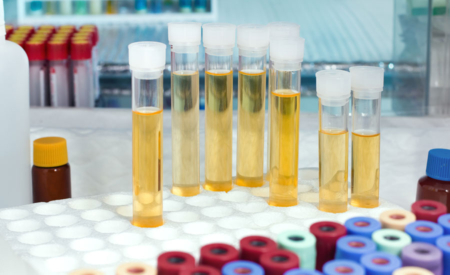
As an old friend in medicine, urine used to be and still is a great companion for diagnostics: Here is why
Urine metabolomics in short
Urine metabolomics offers a particularly interesting readout of the metabolic status of an organism for 3 main reasons:
- It can be collected non-invasively
- It is produced regularly and in large quantities
- It harbors a wealth of information on the body’s metabolic activity
To maximize the potential of a urine metabolomics study, attention should be given to the following steps:
- Study design, time of collection, number of time points
- Sample collection, processing and storage
- Reproducibility and quantitative measurements
- Robust data analysis accounting for urine-specific confounders
Biocrates has produced an Application Note dedicated specifically to the measurement of urine samples with targeted metabolomics kits.
If you want to know more about urine, its uses in medicine and research and the technical aspects that should be considered when planning and conducting such experiments, keep reading our blog below.
Urine – An old friend of medicine

The analysis of urine has been part of the medical toolkit for thousands of years. Long before the dawn of modern analytical methods, the color, smell, and even taste of urine were used to diagnose diverse ailments ranging from kidney disease to diabetes (Armstrong 2007). Since urine is produced by filtration of the blood by the kidneys, it is no surprise that it is often used to study kidney function. However, urine is also a rich source of information on the functionality of other organs at the metabolic level, making it valuable for a broad range of biological investigations.
Urine is a material of choice for medical and biological research, primarily for three reasons:
- It can be collected non-invasively
- It is produced regularly and in large quantities
- It contains a wealth of information related to the body’s metabolic activity
In this article, we will discuss the applicability of urine to omics, its usefulness for biomarker discovery and mechanistic research, and the specific challenges and opportunities related to its use for metabolomics.
Urine composition and omics
Urine has been demonstrated to be a valuable sample matrix across multiple omics fields. Transcriptomics has provided a wealth of information on mRNAs, miRNAs, and other non-coding RNAs, both cell-free and from exfoliated cells present in the urine (Kim et al. 2020).
Under physiological conditions, urine contains low levels of proteins since they are mostly retained in the blood after renal filtration. Nevertheless, the urinary proteome has proven to be rich in information, even in healthy individuals (Adachi et al. 2006; Marimuthu et al. 2011). Lipidomics (the sub-branch of metabolomics focused on lipids) is also being carried out on urine samples. Although circulating lipids are mostly retained in the blood, lipidomics has already provided promising results (Graessler et al. 2018). In contrast, urine is characterized by high levels of metabolites from various small molecule classes, including “metabolic waste” products brought by the circulation to the kidneys for excretion. These constitute a wealth of information for metabolomics that pertains to both renal function and metabolic processes ongoing in the body.
Urine and targeted metabolomics
Biocrates was one of the first contributors to the Urine Metabolome Database with measurements from the AbsoluteIDQ® p180 kit (Bouatra et al. 2013) . With this kit, 57 metabolites representing 5 classes (amino acids, amino acid related, biogenic amines, nucleobases and related and acylcartinines) are consistently found in human urine (Application Note). Biocrates has since launched the MxP® Quant 500 kit, featuring a more comprehensive metabolite coverage to quantify up to 111 metabolites (mostly small molecules, but also a few lipids) from the following 17 classes that are consistently found in human urine:
- Alkaloids
- Amine oxides
- Amino acids
- Amino acid related
- Bile acids
- Biogenic amines
- Carboxylic acids
- Cresols
- Fatty acids
- Hormones & related
- Indoles & derivatives
- Nucleobases & related
- Vitamins & cofactors
- Acylcarnitines,
- Cholesteryl esters
- Diglycerides
- Triglycerides
For more detail on the metabolites detected in human urine with Biocrates’ kits, see our Application Note.
Urine metabolomics as a source of biomarkers
Disease biomarkers are greatly sought after in the diagnostic world. Finding biomarkers in biofluids that can easily be obtained from patients in large volumes such as urine presents an ideal scenario from a collection and analysis standpoint. Urine metabolomics has already proven an effective means to identify biomarker candidates for bladder cancer (Wittmann et al. 2014), interstitial cystitis (Parker et al. 2016), esophageal squamous cell carcinoma (Xu et al. 2016), inflammatory bowel disease (Dawiskiba et al. 2014) and more.
Given how easy it is to collect urine samples, it might be tempting to plan a quick metabolomics experiment to evaluate the potential of the method for one’s research. However, in order for the results to really discriminate between groups upon statistical analysis, several important points must be taken into consideration to avoid losing statistical power to unforeseen confounders.
Technical considerations
Urinary volume and flow rate vary greatly between individuals and even for one individual throughout the day. This is because, in addition to their blood filtering and secretion functions, the kidneys also adjust the volume and pH of the blood by modulating the volume of water and concentrations of inorganic salts excreted into the urine. These peculiarities and other technical considerations are discussed in the paragraphs below.
Normalization of absolute metabolite concentrations
The absolute concentrations of metabolites in the urine (by definition relative to the volume of the sample) can vary greatly, even for a single healthy individual. Thus, when performing absolute quantifications in urine samples (as is the case with all of Biocrates’ products) comparison between samples is made difficult if no normalization is applied. This is classically compensated for by applying a normalization to urinary creatinine levels. Creatinine is already included in all our broad panel metabolomics kits.
Ion suppression effect
The salts and metabolites that are filtered out of the blood and/or secreted into the urine can cause an ion suppression effect that interferes with mass spectrometry measurements (Annesley 2003). For this reason, urine samples are often diluted before measurement. The ratio for dilution (usually 1:5 to 1:20) depends on the initial dilution of the urine sample, which itself depends on the level of hydration of the subject, among other factors. For this reason, it is advisable to directly measure one or more dilutions of each sample in the same run to avoid having to re-pipet an entire experiment.
Sample collection, processing and storage
Most of the rules that apply to the collection and handling of other biological material also apply to urine. Below are some of the important points to keep in mind:
- Handle samples promptly: in order to limit sample degradation and contamination, place samples at 4°C shortly after collection (Roux et al. 2015).
- Do not assume that the samples are sterile: it is now commonly accepted that the legendary sterility of urine is compromised as soon as the fluid leaves the urethra
- Plan experiments thoroughly: factors such as the time of day, fasting status, or even diet can strongly influence metabolism and in turn what will be present in the urine
- Collect without preservative (if possible): unlike blood, urine does not require the use of collection tubes containing a preservative. However, some labs choose to use additives to avoid bacterial growth, which can influence metabolomic readouts.
- Apply mild centrifugation promptly and before freezing: pellet and remove the cells that are normally exfoliated into the urine. If the cells are not removed or if the centrifugation is too harsh and breaks their plasma membranes, their intracellular contents are released and might alter the results.
- Filter before freezing (optional): filtration can help remove additional cell debris but at the risk of filtering out some of the metabolites.
- Keep freeze-thaw cycles to a minimum: these can dramatically influence metabolite stability. A maximum of 2 freeze-thaw cycles is recommended (Rotter et al. 2017).
For more references and recommendations on how to collect and prepare urine samples for metabolomics, see the White Paper on preanalytical processing of biological samples for metabolomics by Kirwan et al. (2018) or contact us.
Links
Related products
MxP® Quant 500 kit, AbsoluteIDQ® p180 kit, AbsoluteIDQ® p400 HR kit, Quant HR XPress™ kit, Services
References
Adachi, Jun; Kumar, Chanchal; Zhang, Yanling; Olsen, Jesper V.; Mann, Matthias (2006): The human urinary proteome contains more than 1500 proteins, including a large proportion of membrane proteins. In Genome Biology 7 (9), R80. DOI: 10.1186/gb-2006-7-9-r80.
Annesley, Thomas M. (2003): Ion suppression in mass spectrometry. In Clinical chemistry 49 (7), pp. 1041–1044. DOI: 10.1373/49.7.1041.
Armstrong, J. A. (2007): Urinalysis in Western culture: a brief history. In Kidney international 71 (5), pp. 384–387. DOI: 10.1038/sj.ki.5002057
Bouatra, Souhaila; Aziat, Farid; Mandal, Rupasri; Guo, An Chi; Wilson, Michael R.; Knox, Craig et al. (2013): The human urine metabolome. In PloS one 8 (9), e73076. DOI: 10.1371/journal.pone.0073076.
Dawiskiba, Tomasz; Deja, Stanisław; Mulak, Agata; Ząbek, Adam; Jawień, Ewa; Pawełka, Dorota et al. (2014): Serum and urine metabolomic fingerprinting in diagnostics of inflammatory bowel diseases. In World journal of gastroenterology 20 (1), pp. 163–174. DOI: 10.3748/wjg.v20.i1.163
Graessler, J.; Mehnert, C. S.; Schulte, K-M; Bergmann, S.; Strauss, S.; Bornstein, T. D. et al. (2018): Urinary Lipidomics: evidence for multiple sources and sexual dimorphism in healthy individuals. In The pharmacogenomics journal 18 (2), pp. 331–339. DOI: 10.1038/tpj.2017.24.
Kim, Jayoung; Kim, Won Tae; Kim, Wun-Jae (2020): Advances in urinary biomarker discovery in urological research. In Investigative and clinical urology 61 (Suppl 1), S8-S22. DOI: 10.4111/icu.2020.61.S1.S8.
Kirwan, Jennifer A.; Brennan, Lorraine; Broadhurst, David; Fiehn, Oliver; Cascante, Marta; Dunn, Warwick B. et al. (2018): Preanalytical Processing and Biobanking Procedures of Biological Samples for Metabolomics Research: A White Paper, Community Perspective (for “Precision Medicine and Pharmacometabolomics Task Group”-The Metabolomics Society Initiative). In Clinical chemistry 64 (8), pp. 1158–1182. DOI: 10.1373/clinchem.2018.287045.
Marimuthu, Arivusudar; O’Meally, Robert N.; Chaerkady, Raghothama; Subbannayya, Yashwanth; Nanjappa, Vishalakshi; Kumar, Praveen et al. (2011): A Comprehensive Map of the Human Urinary Proteome. In Journal of proteome research 10 (6), pp. 2734–2743. DOI: 10.1021/pr2003038.
Parker, Kaveri S.; Crowley, Jan R.; Stephens-Shields, Alisa J.; van Bokhoven, Adrie; Lucia, M. Scott; Lai, H. Henry et al. (2016): Urinary Metabolomics Identifies a Molecular Correlate of Interstitial Cystitis/Bladder Pain Syndrome in a Multidisciplinary Approach to the Study of Chronic Pelvic Pain (MAPP) Research Network Cohort. In EBioMedicine 7, pp. 167–174. DOI: 10.1016/j.ebiom.2016.03.040
Rotter, Markus; Brandmaier, Stefan; Prehn, Cornelia; Adam, Jonathan; Rabstein, Sylvia; Gawrych, Katarzyna et al. (2017): Stability of targeted metabolite profiles of urine samples under different storage conditions. In Metabolomics : Official journal of the Metabolomic Society 13 (1), p. 4. DOI: 10.1007/s11306-016-1137-z.
Roux, Aurélie; Thévenot, Etienne A.; Seguin, François; Olivier, Marie-Françoise; Junot, Christophe (2015): Impact of collection conditions on the metabolite content of human urine samples as analyzed by liquid chromatography coupled to mass spectrometry and nuclear magnetic resonance spectroscopy. In Metabolomics : Official journal of the Metabolomic Society 11 (5), pp. 1095–1105. DOI: 10.1007/s11306-014-0764-5.
Wittmann, Bryan M.; Stirdivant, Steven M.; Mitchell, Matthew W.; Wulff, Jacob E.; McDunn, Jonathan E.; Li, Zhen et al. (2014): Bladder cancer biomarker discovery using global metabolomic profiling of urine. In PloS one 9 (12), e115870. DOI: 10.1371/journal.pone.0115870.
Xu, Jing; Chen, Yanhua; Zhang, Ruiping; He, Jiuming; Song, Yongmei; Wang, Jingbo et al. (2016): Global metabolomics reveals potential urinary biomarkers of esophageal squamous cell carcinoma for diagnosis and staging. In Scientific Reports 6, p. 35010. DOI: 10.1038/srep35010

